World Journal of Pharmaceutical
and Medical Research

( An ISO 9001:2015 Certified International Journal )

An International Peer Reviewed Journal for Pharmaceutical and Medical Research and Technology
An Official Publication of Society for Advance Healthcare Research (Reg. No. : 01/01/01/31674/16)
ISSN 2455-3301
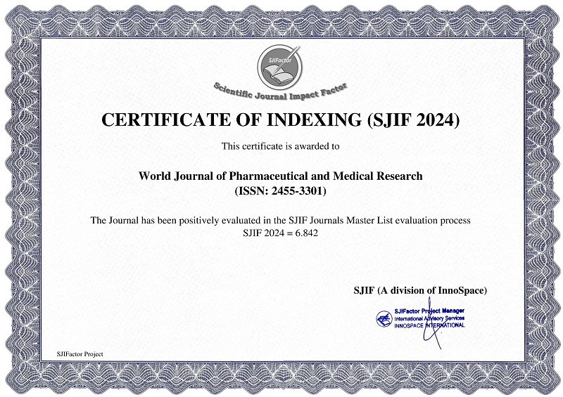
IMPACT FACTOR: 6.842

ICV : 78.6

World Journal of Pharmaceutical and Medical Research (WJPMR) has indexed with various reputed international bodies like : Google Scholar , Index Copernicus , SOCOLAR, China , Indian Science Publications , Cosmos Impact Factor , Research Bible, Fuchu, Tokyo. JAPAN , Scientific Indexing Services (SIS) , UDLedge Science Citation Index , International Impact Factor Services , International Society for Research Activity (ISRA) Journal Impact Factor (JIF) , International Innovative Journal Impact Factor (IIJIF) , Scientific Journal Impact Factor (SJIF) , Global Impact Factor (In Process) , Digital Online Identifier-Database System (DOI-DS) , Science Library Index, Dubai, United Arab Emirates , Eurasian Scientific Journal Index (ESJI) , International Scientific Indexing, (ISI) UAE , IFSIJ Measure of Journal Quality , Web of Science Group (Under Process) , Directory of Research Journals Indexing , Scholar Article Journal Index (SAJI) , International Scientific Indexing ( ISI ) , Scope Database , Academia , 

Abstract

EVALUATION OF LUNG MASSES USING COMPUTED TOMOGRAPHY AND COMPARASION OF RESULTS WITH HISTOPATHOLOGY.

Dr. Shabir Ahmad Bhat, Dr. Shadab Maqsood and *Dr. Iqubal Hussain Dar

ABSTRACT

This prospective study conducted in the tertiary care hospital setting included 50 subjects(31males,19 females)in the age group of 20-80(mean,54±15.43)years with radiological evidence of parenchymal lung shadows. These cases were subjected to detailed history, clinical examination and laboratory investigations. The radiographic features were confirmed by computed tomography (CT) and histopathology. Anaemia and elevated ESR were noted in a significant proportion of cases .Majority of the masses involved the right upper lobes; 64% of the malignant masses had ill-defined or irregular margins and 54% masses were 4 to 6 cm in size. CT examination picked up findings not appreciable on chest radiography .We conclude that CT proves to be a better diagnostic modality to determine the location, size and character of the mass shadows, and helps significantly in differentiating benign from the malignan lesions.

[Full Text Article]    [Download Certificate]

Powered By WJPMR | All Right Reserved

WJPMR